The Pediatric PICC Placement Simulator is a high-fidelity 3D-printed training model designed for
ultrasound-guided placement of peripherally inserted central PICC in children.
Developed from CT scan of real pediatric patients, the phantom reproduces the key
anatomical structures of the upper limb, including the humerus, brachial artery, basilic vein,
brachial veins, cephalic vein and surrounding soft tissues.
PICC placement in pediatric patients is a delicate procedure requiring high precision. This realistic simulator provides a safe platform for hands-on practice, boosting trainee confidence and coordination without any patient risk.
-
Anatomical Realism: Derived from real pediatric CT scans to preserve accurate dimensions and spatial relationships between vessels, soft tissues, and bone landmarks.
-
Realistic Ultrasound Visibility: Internal structures replicate clinical-grade ultrasound visibility for accurate vessel identification.
-
Puncture Simulation: Covers the entire Seldinger technique—from ultrasound exploration and vein puncture with tactile feedback to fluid reflux confirmation and guidewire insertion.
-
Safe & Repeatable Practice: Eliminates clinical risks while supporting repeated punctures, allowing trainees to master the procedure through continuous refinement.
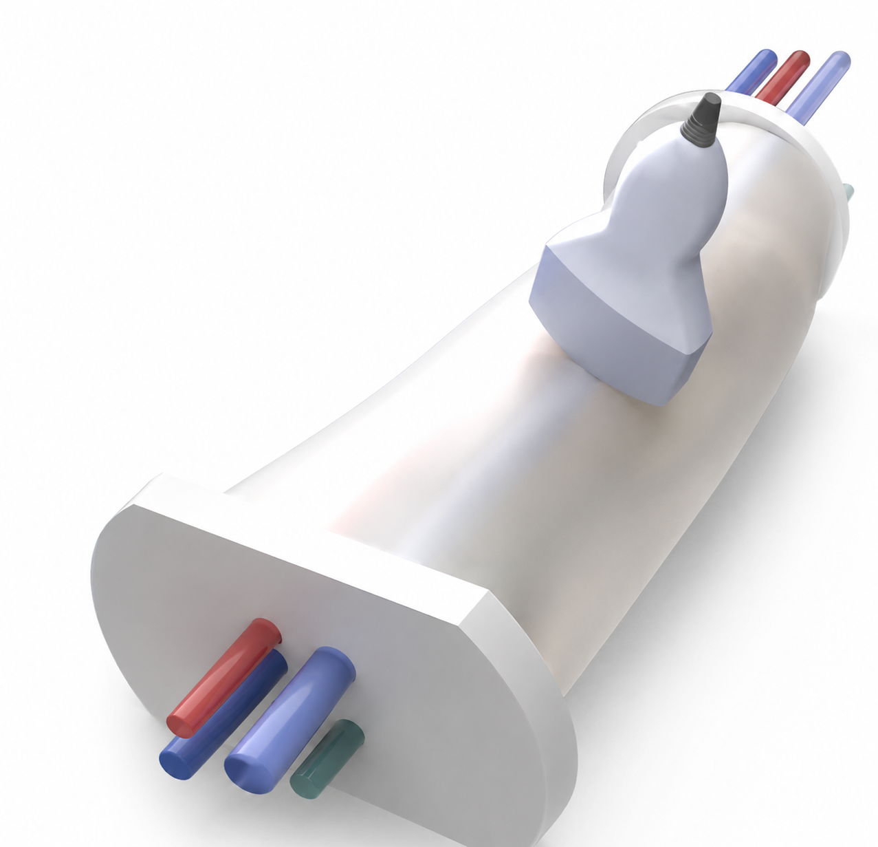
The simulator is shipped ready to be used, comprehensive digital and paper instructions, along with video tutorials, are provided upon simulator delivery.
Looking for a customized version?
The simulator can be customized with specific inflow- outlflow connectors pathological conditions or uncommon anatomies. Customizations can be
requested at info@3d4med.eu.