One of the most delicate cases managed at the Niguarda center dedicated to heart disease during pregnancy in recent years.
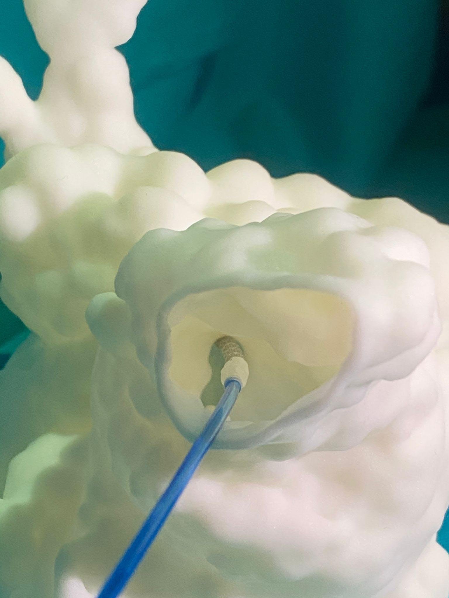
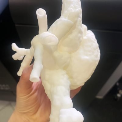
This is the story of Tamara, 34 years old, who wanted to have a baby and managed to make her dream come true despite a delicate heart surgery performed during pregnancy. The testing in preparation for the surgery – at the Milan hospital – was done on a “digital twin” of Tamara’s heart, a 3D printed model of the organ. The “twin heart” – reconstructed and produced by 3D4Med Clinical 3D Printing Laboratory – reproduces the real one in every detail.
Tamara was born with a condition called transposition of the great arteries, where the aorta and pulmonary artery are reversed, resulting in an abnormal anatomy.
The clinical specialist asked for a replica of Tamara’s heart, produced by means of 3D printing technology, to be used for training before the surgery in order to know in advance all the steps of the procedure and to choose the most suitable instrumentation. The medical staff of the hospital and the engineers of the “3D4Med Clinical 3D Printing Lab” of the Fondazione IRCCS Policlinico San Matteo of Pavia teamed up with the cardiologists of Niguarda Hospital to produce the three-dimensional model, reconstructed from the anatomical images acquired by the cardiac magnetic resonance experts of the Niguarda. The surgery was performed in the 16th week of pregnancy. Jacopo Oreglia, head of hemodynamics and interventional cardiology, explains: “The tests carried out allowed us to speed up the procedure and about 30 minutes of irradiation were enough for the X-ray navigation system, which was also used at minimum power, to limit the risks for the child “, born in the 31st week by cesarean section.
After a few weeks, mom and baby returned home. Heart surgery during pregnancy was successful !
Abnormal anatomy

Virtual model

Surgical simulation


