Abdominal Surgery: Planning & Simulation
Our 3D printed models are widely used at IRCCS Policlinico San Matteo of Pavia to plan several surgical procedures, especially involving pancreas, spleen and kidney. Most of surgical interventions are performed by means of laparoscopic or robotic procedures.
3D Printed Models
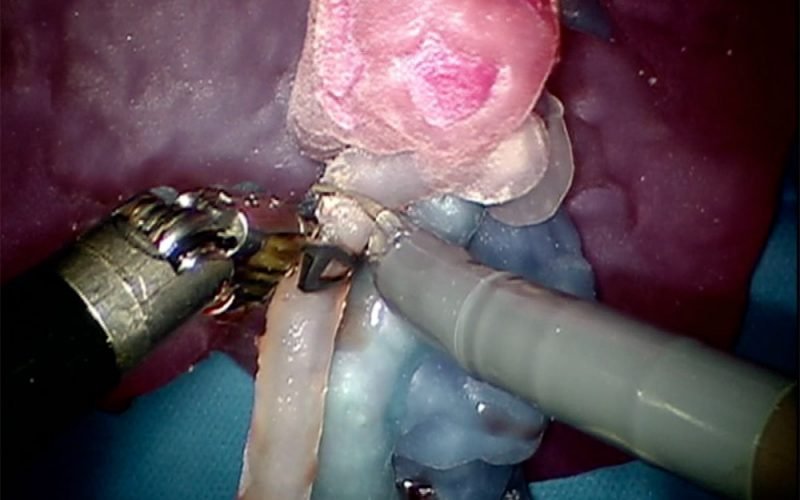
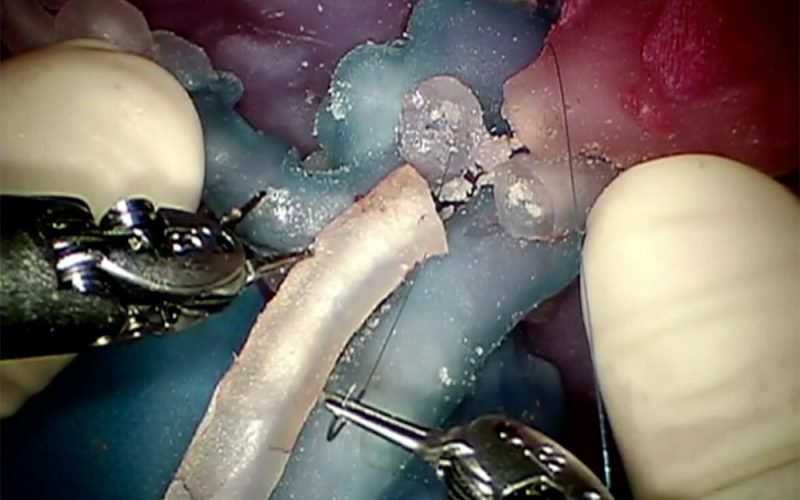
Using an Objet260 Connex3, 3D models can be printed in colored and deformable resins. This technology enables the tuning of deformability and transparency features, allowing to replicate as much as possible real abdominal organs and vessels, both in morphological and mechanical properties. These deformable models can be useful for surgical simulations performed through DaVinci Robotic Surgical System®: the surgeon performs the surgery directly on the model, anticipating and preventing risks and problems related to the surgical procedure under investigation.
Using a Projet460 Plus, plaster 3D printed models with a full-scale chromatic resolution can be produced to help surgeons in surgical planning and as an intra-operative guide, allowing an effective chromatic discrimination of all the involved anatomical structures.
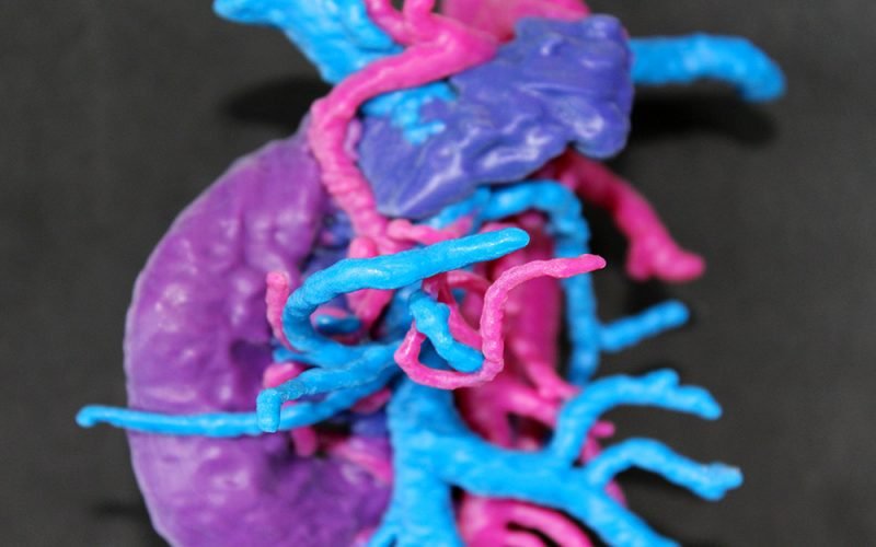
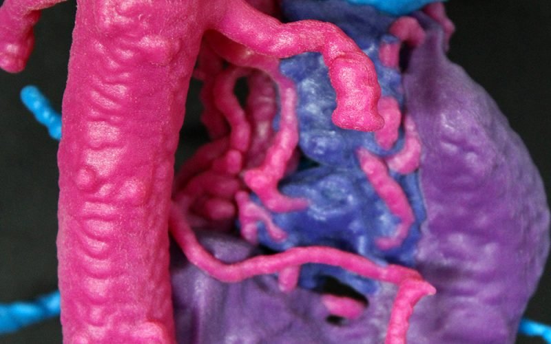
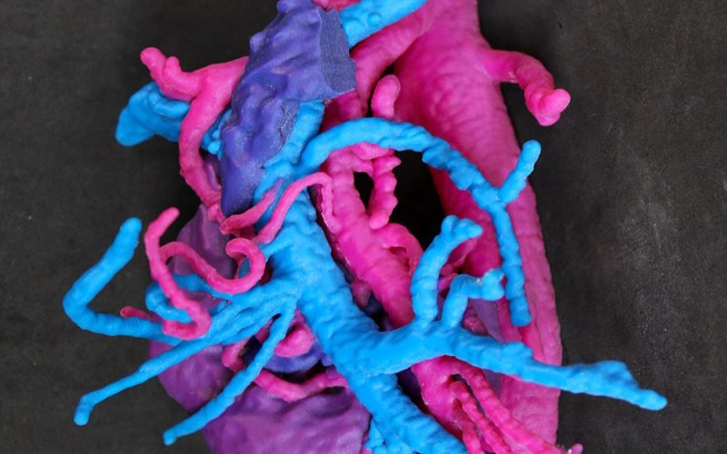
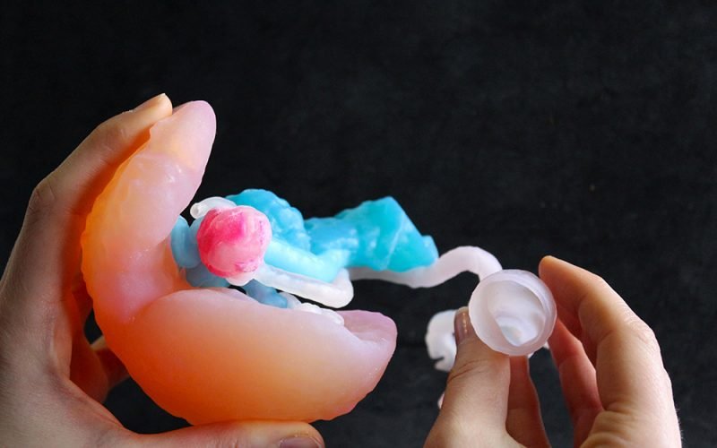
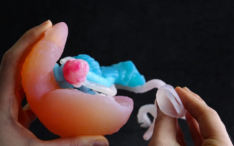
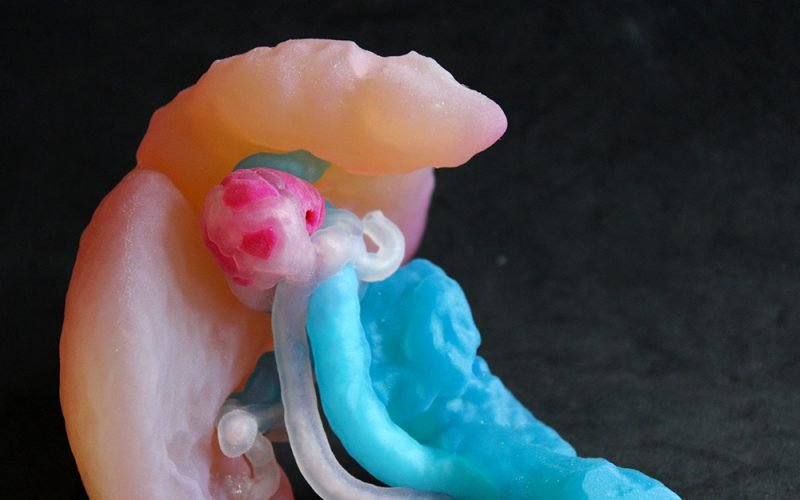
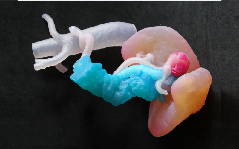
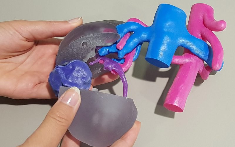
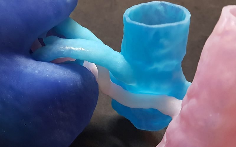
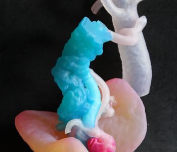
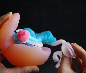
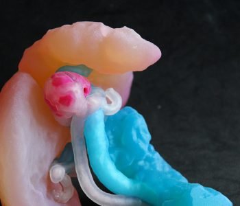
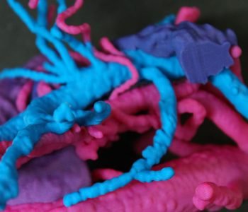
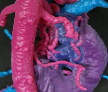
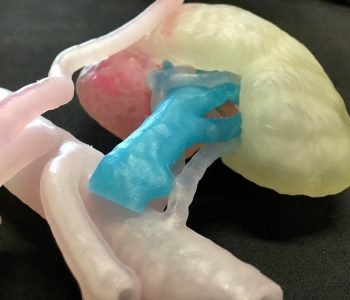
The 3D printed patient-specific model represents a case of splenic artery affected by an aneurysm in the proximal tract, near the spleen. The spleen, the main arterial and venous afferent blood vessels, the splenic aneurysm and the pancreas have been reconstructed. The model has been printed in colored and deformable resins. Especially vessels are reconstructed and 3D printed in order to replicate as much as possible real vessels, both in morphological and mechanical properties. This allowed the use of the model for surgical simulation performed through DaVinci Robotic Surgical System®.
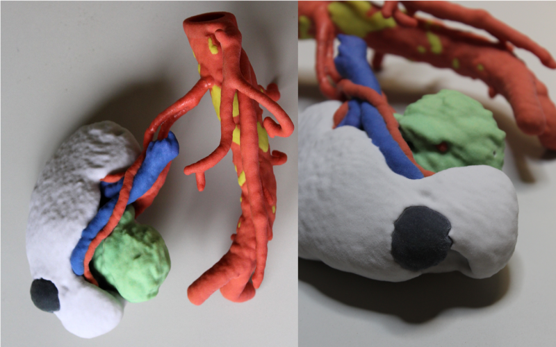
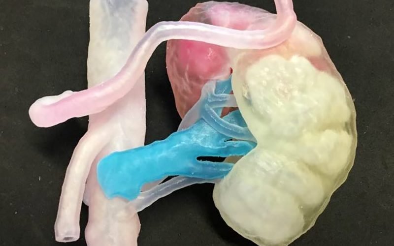
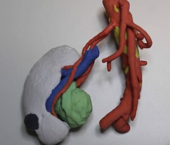
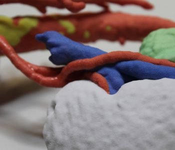
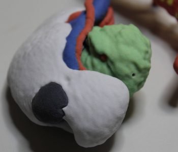
The patient-specific model represents a kidney (white) with its tumor (black), a great renal cyst (green) and the surrounding venous (blue) and arterial (red) vessels with some calcification (yellow) on the aorta. The model has been used to properly plan the surgical resection of the kidney tumor and cyst performed through DaVinci Robotic Surgical System®. With this model the surgeon could appreciate the morphology of the vessels and the relationships between all the anatomical and pathological structures. The clinical case is particularly interesting due to an anomalous conformation of the arterial vessels afferent to the kidney.
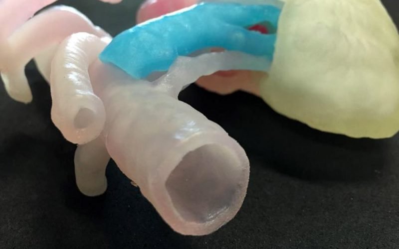
The patient-specific model represents the pancreatic-duodenal tract with its arterial and venous vessels. The model was useful for the surgical planning of an intervention of total mesocolic excision in right hemicolectomy. The pancreatic-duodenal tract is characterized by a particularly complex vasculature: the 3D printed model has allowed a better and easier visualization of the vessel’s structure both before and during the robotic intervention.
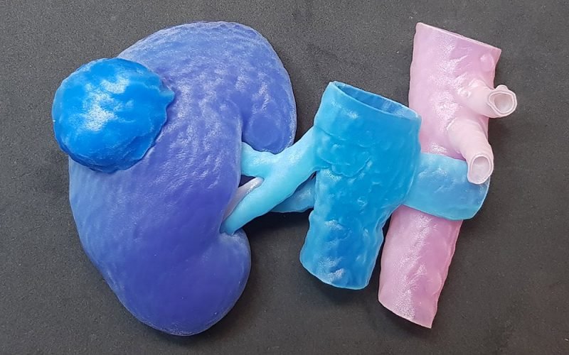
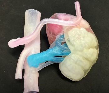
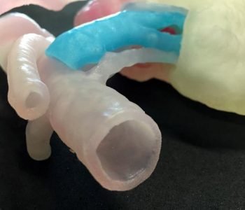
The model represents the kidney of a living donor to be procured for a kidney transplant. The model has been printed in colored and deformable resins, to allow the surgical simulation of the intervention performed through DaVinci Robotic Surgical System®. Particular attention is paid to the reproduction of vessels, with the aim to reproduce with the highest realism both morphological and mechanical properties.
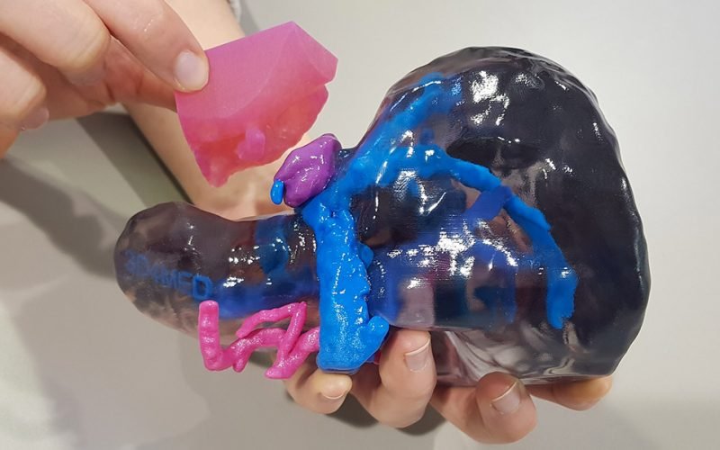
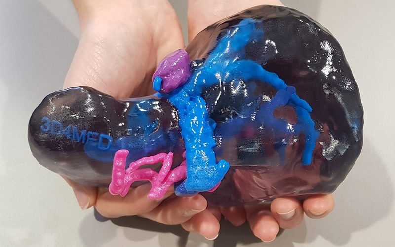
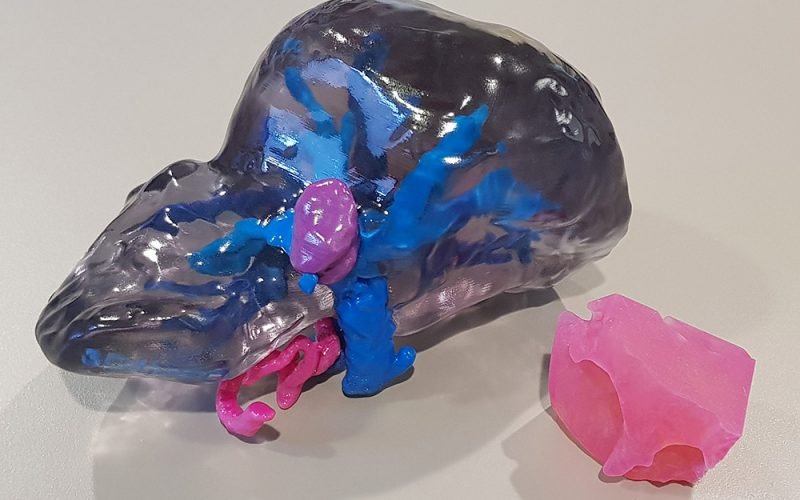
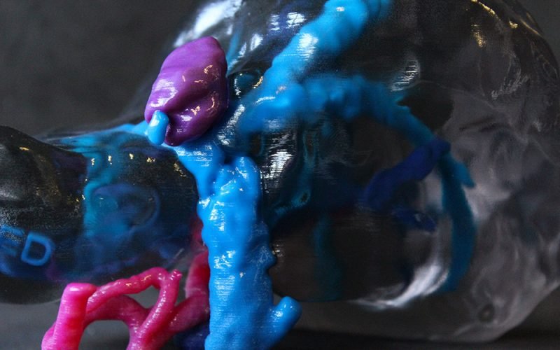
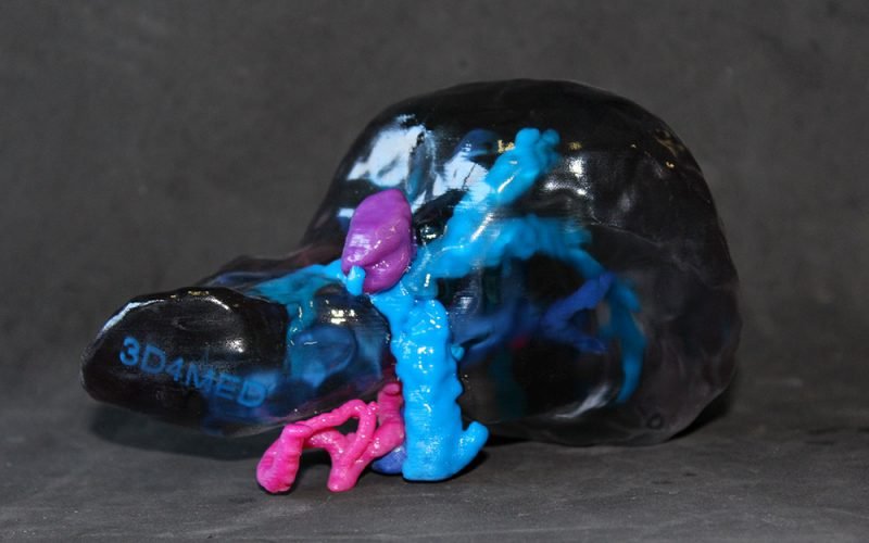
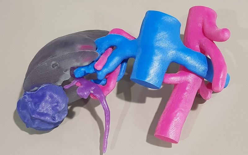
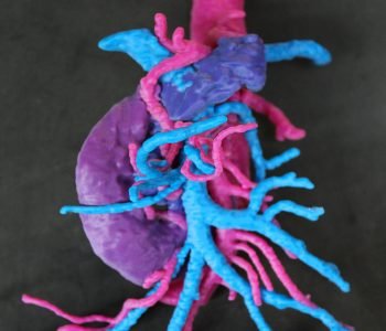
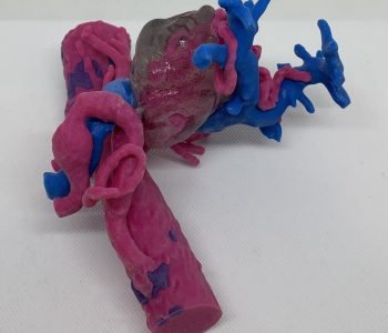
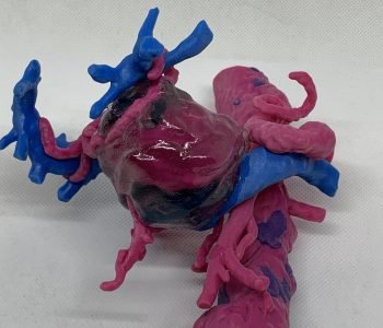
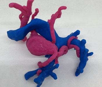
The patient-specific model represents the hepatic vascular tract, including both arteries (magenta) and veins (blue); the hepatic artery is affected by an extensive aneurysm, also presenting a thrombotic part (transparent). A second model – of the same clinical case – was also printed without the thrombus, to better see and evaluate the vascular structures near to the aneurysm.
After models’ evaluation, surgeons decided to not proceed with the intervention for the aneurysm exclusion. He was then observed by an interventional radiologist, in order to evaluate a stent positioning in the hepatic aneurysm site.


