Urology
3D printed models support the urologist in the surgical planning of renal intervention
The use of 3D printing allows to obtain an accurate replica of the patient part, the pre-operative planning is an essential phase of the surgery, and sometimes the only way to carry out a highly complicated intervention.
The use of 3D printing allows to obtain an accurate replica of the patients part, with the possibility to finely adjust the incisions and to test different instruments, inserting them in the same places where they will be used in the surgery.
The patient underwent a laparoscopic intervention for kidney tumor enucleation; the surgery was performed following a selective clamping procedure of the left renal artery with fluorescence for the selective ischemia of the kidney parenchyma next to the tumor mass.
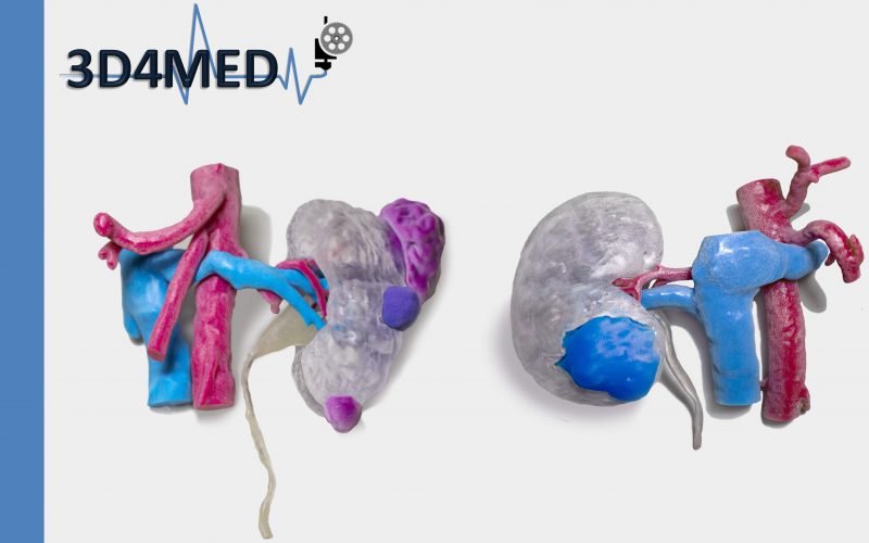
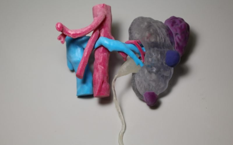
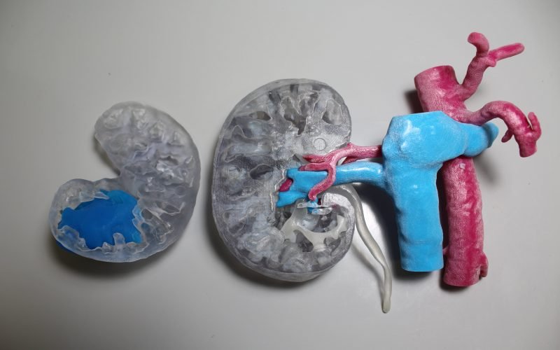
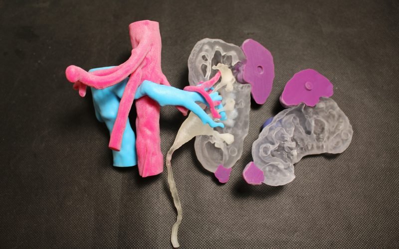
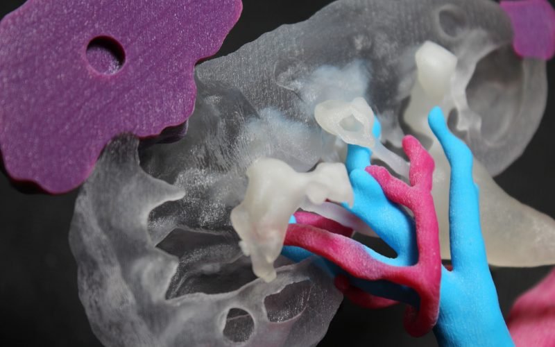
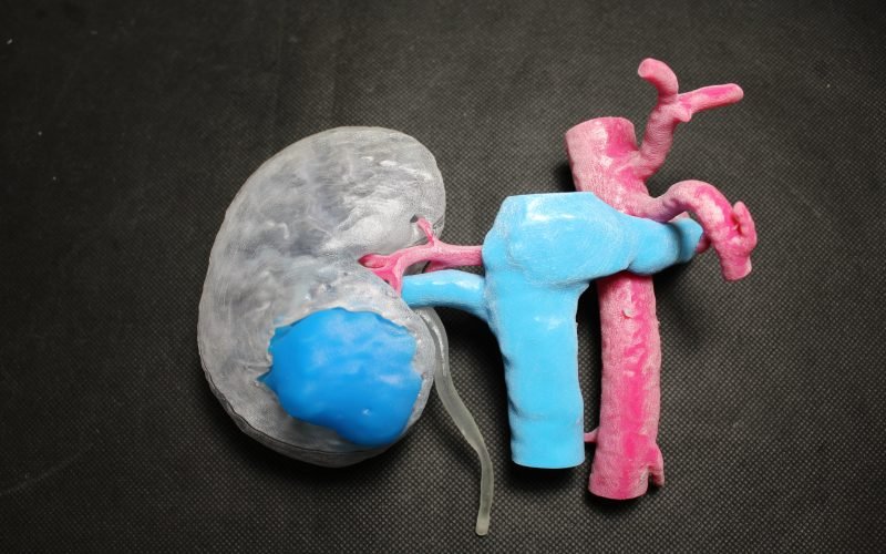
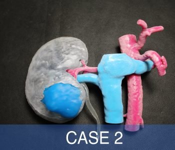
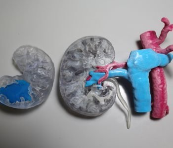
The 3D printed models involve different anatomical structures of interest, printed with the appropriate photopolymer resins.
The model is made up of interlocking parts, enabling the assessment of the infiltration level of the tumor and an eventual (not occurred) involvement of blood vessels or ureter.
The clinical case will be presented as a video case report to the Asian Urology WebCongress taking place this month.
As the previously described clinical case, the 3D printed model is produced with interlocking parts and involves both arterial and venous blood vessels, the ureter, the tumor mass, and the kidney parenchyma. The same materials of Case 1 are here employed, thus with a deformable urether. Here the tumor has significantly larger dimensions, it infiltrates deeper in the kidney parenchyma and it extends very close to the arterial and venous vessels and the ureter.
The 3D printed model will be used as a didactic tool to improve the anatomical comprehension and the study of the degree of infiltration of kidney tumors’.


