Interventional radiology
3D printed models are useful for the morphological evaluation of aneurysms and for the planning / simulation of endoprostheses’ positioning.
3D printed models for the planning and simulation of stent placement for the treatment of aneurysms involving the peripheral arterial system. The models have been helpful in the selection of the proper stent size to restore the right blood flow in both single-sided and bilateral renal aneurysms. Moreover. the use of deformable resins allows direct stimulation of the device positioning on the model.
3D printed model for the evaluation of the proper interventional approach for the treatment of a renal arterial aneurysm. The model showed the presence of an unusual angle of the renal artery due to the external compression from the diafframatic pillar.
Forensic medicine
3D printed models support the medical examiner in the analysis of the findings.
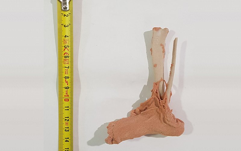
Upon assignment by the Judicial Authority, these finds were subjected to technical-specialist investigations to determine if they were of human origin.
The morphological peculiarities of exhibit A have requested the execution of radiological insights. A thin section of fabric was made of bone from a fragment of the tibia, for microscopic observation. In the end, a reproduction was made in plaster with the 3D printer (thanks to the collaboration with the 3D Printing Lab for Surgery of 3D4MED), to facilitate the comparisons with collections of skeletons at museums of human and veterinary anatomy.
Genetic diseases
The 3D printed model is useful for the choice and planning of the therapeutic/surgical approach in the case of very rare and complex malformations/genetic diseases.
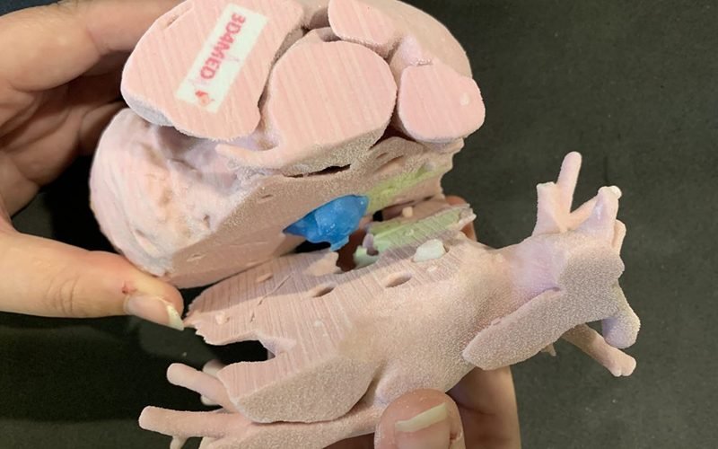
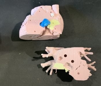
Model for the clinical evaluation of an extremely rare congenital pathology: paediatric case of 3 cardiac mixoma
Model made of interlocking parts produced with different 3d printing technologies to allow a deeper visualization and comprehension of anatomical and pathological structures.


