Cardiology
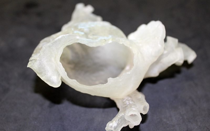
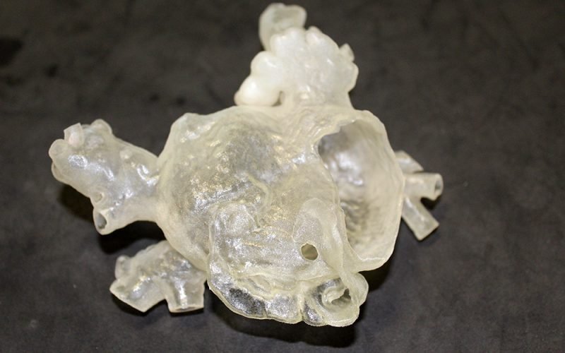
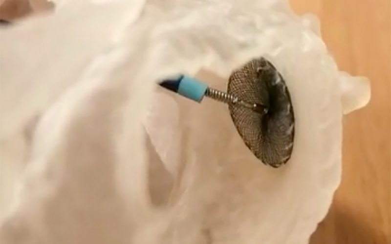
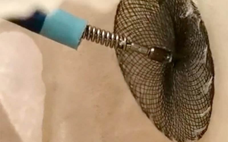
3D printing provides a concrete and precise reproduction of the morphological and pathological aspects of the heart chambers, allowing the surgeon to plan and simulate interventional procedures choosing the most suitable device.
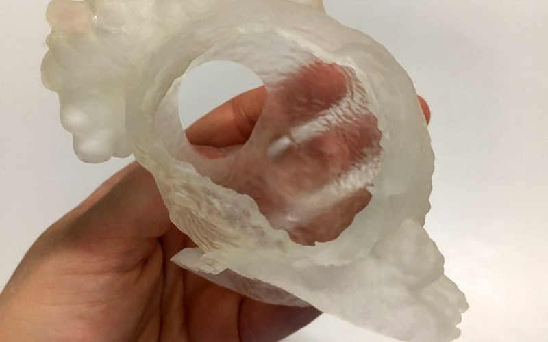
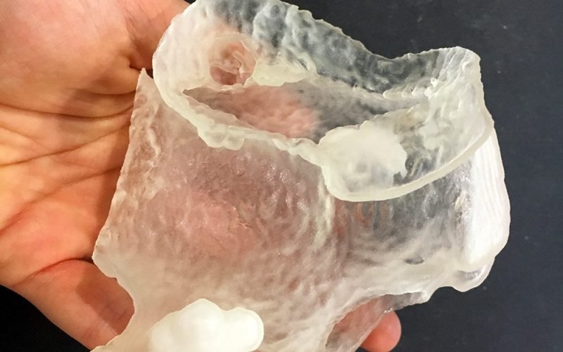
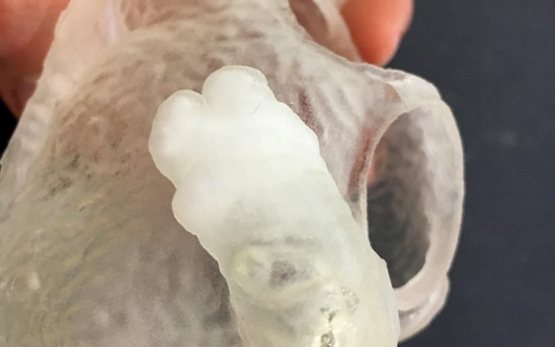
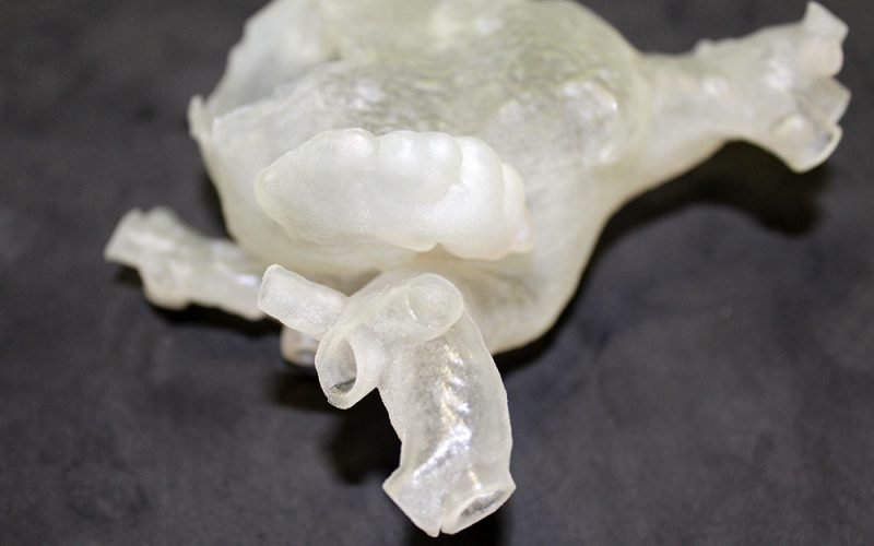
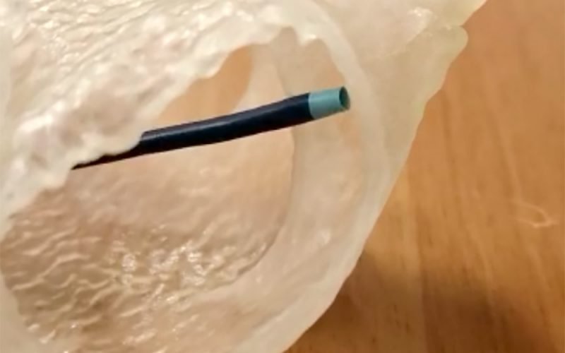
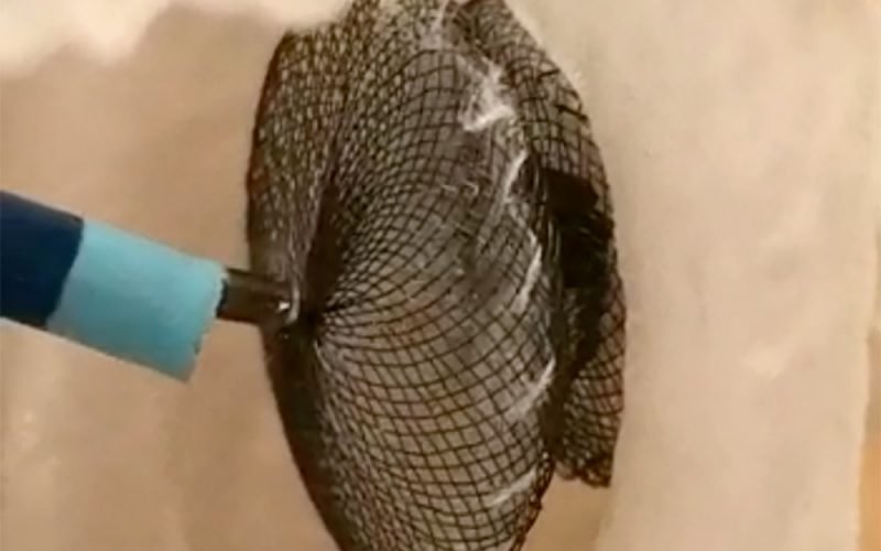
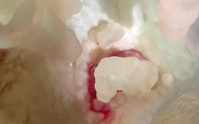
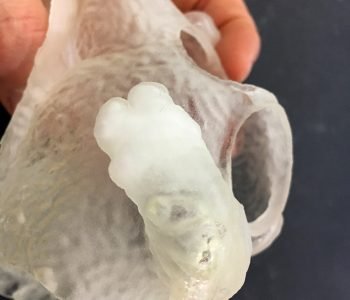
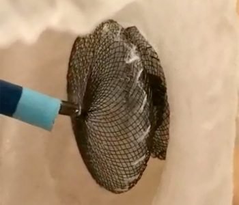
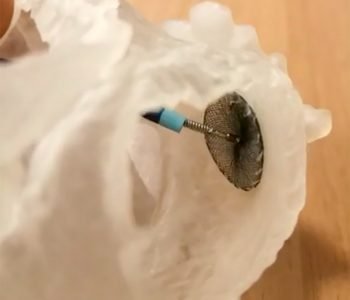
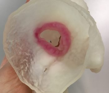
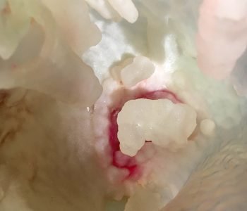
The 3D printed model describes a left atrium with a “chicken wing” appendage. The model has been printed in deformable material faithfully mimicking the atrial wall mechanical behaviour. The model has been useful to assess trans-catheter closure appendage feasibility and to plan the procedure. The anomalous anatomy of the patient increased the technical complexity of the intervention: thus, the procedure’s simulation on the 3D printed model has helped the surgeon in the choice of the most suitable device.
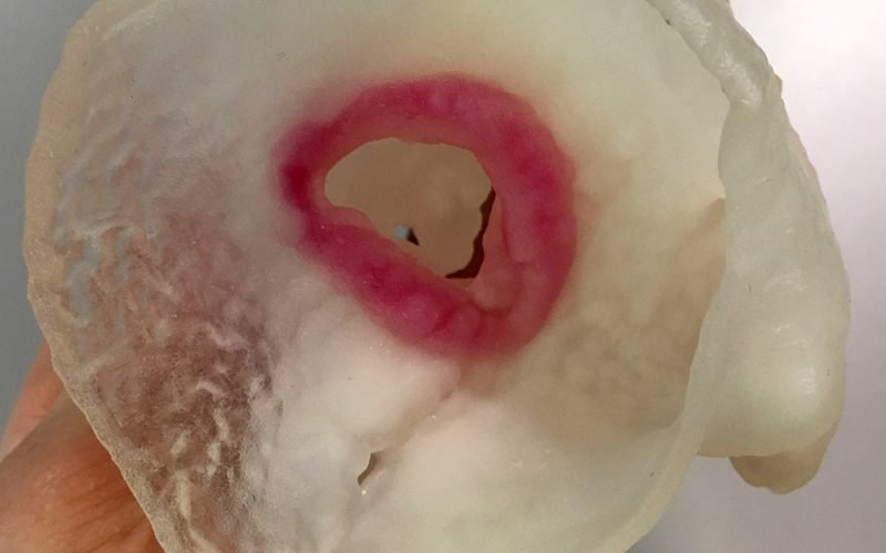
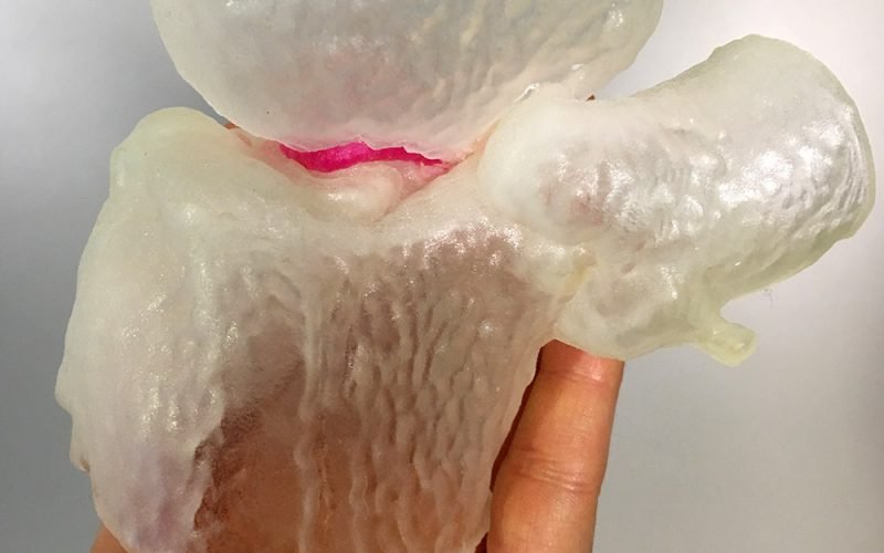
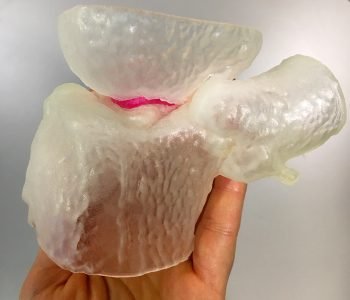
The patient-specific model describes the area between the left atrium and ventricle with its protrusions, the atrial appendage and the first tract of the proximal ascending aorta. The patient has an annular endo-prosthesis already implanted in the atrioventricular junction. The model has been useful to properly plan the placement of a second prosthetic device in the left ventricle. Thanks to the employment of a deformable material it was possible to simulate the procedure directly on the 3D printed model and to select the most suitable device.


