Vascular Surgery
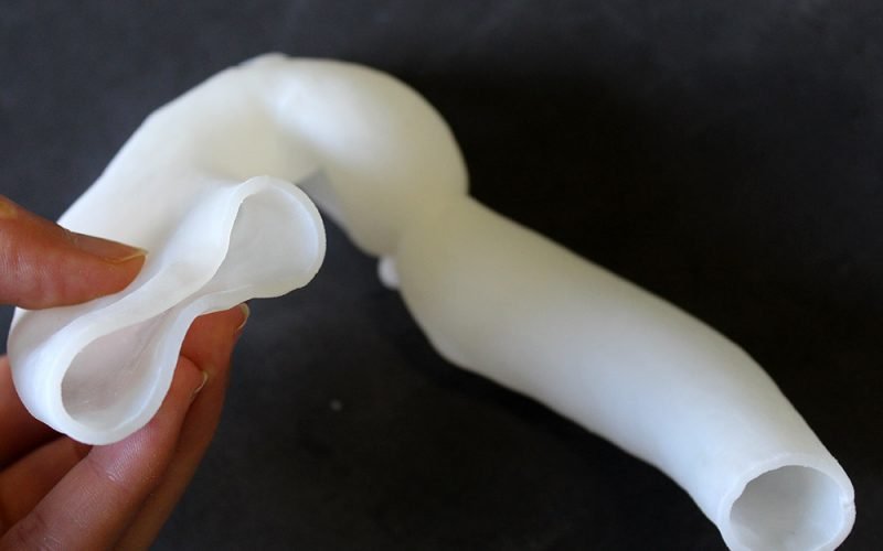
3D printing provides a concrete and precise reproduction of the morphological and pathological aspects of a vascular lesion, allowing the surgeon to plan its proper repair, either by open or endovascular approach.
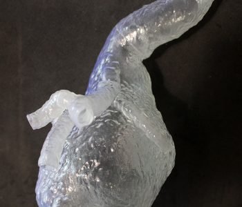
The patient-specific model is a 3D printed reconstruction of an aneurysm of the abdominal Aorta. The model has been useful for the pre-operative evaluation of the aneurysm (shape, dimension and location) and for the surgical planning of aneurysm resection and endoprosthesis selection and placement. The model is printed in transparent photopolymer resin to allow the surgeon to better appreciate the morphology of patient’s vascular anatomy and anomalies and to visualize the positioning of the selected endoprosthesis.
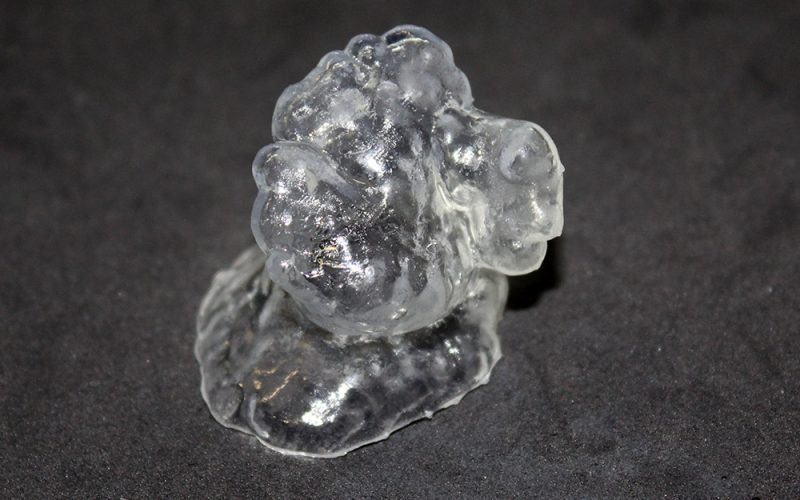
The patient-specific model is a 3D reconstruction of a left Auricle. The left Auricle is a small anatomical structure projecting from the upper anterior portion of the left Atrium. It shows a great inter-patient morphological variability. The 3D printed model includes both the left Auricle and the closest portion of the Atrium. The aim is to allow a better understanding of the specific anatomy and of the right heart malformation’s location with respect to the surrounding anatomical structures and blood vessels. The model has been useful to test different devices in order to select the most suitable for the specific patient.
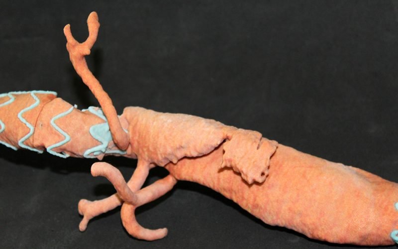
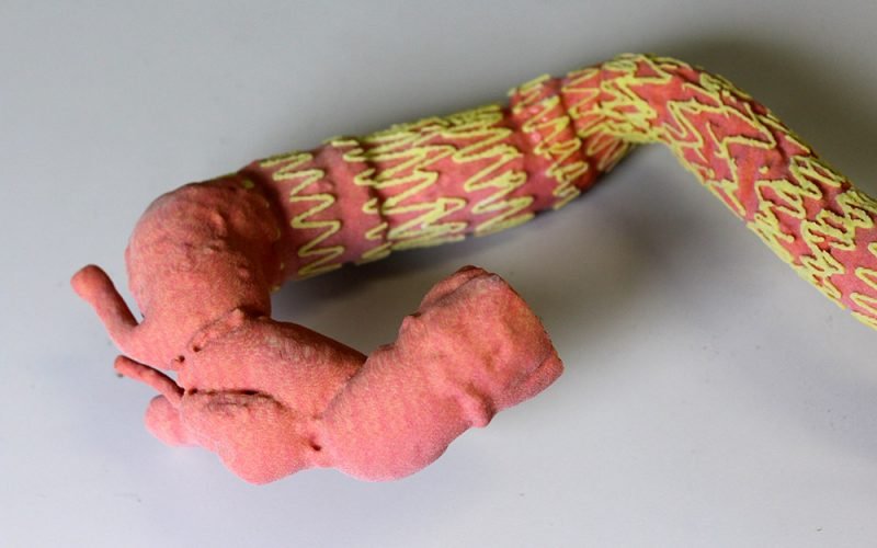
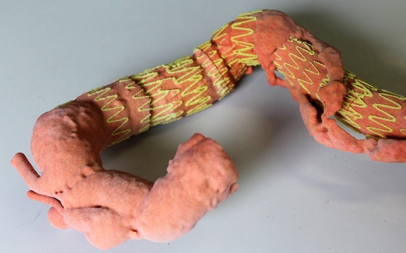
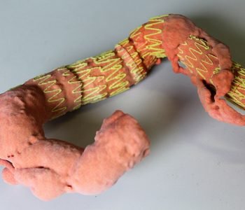
The patient-specific model represents the post-operative scenario of an aortic dissection after stents placement. The model has been useful for the correct endoprosthesis placement evaluation and the monitoring of the course of disease.
The considered patient underwent many surgical interventions and the specific model represents the final outcome after the third endoprosthesis placement.
Since the model is in scale 1:1, its dimensions are significant (about 50 cm length) and it is composed by 3 parts perfectly assembled in post-processing phase.


