Cerebral Artery Aneurysm Clipping: the Potential of 3D Printing
Our 3D printed models helped neurosurgeons to properly plan and simulate cerebral aneurysm clipping procedures. We report here one of the clinical cases treated at Fondazione I.R.C.C.S Policlinico San Matteo of Pavia.
Clinical Case
The patient – diagnosed with a complex brain artery aneurysm – was scheduled for a left middle cerebral artery aneurysm clipping procedure at the bifurcation of M1 and M2, through left pterional craniotomy performed with intraoperative neurophysiological monitoring.
As most of the neurosurgical procedures, aneurysm clipping has several complex and delicate steps to be performed and a careful pre-operative planning could be crucial for the surgery success. Available medical images did not show the aneurysm clearly enough and small mismatches caught by the neurosurgeon from them can turn into larger discrepancies once in the operatory room.
However, our 3D printed model of the patient’s unique anatomy was fundamental to overcome these issues.
How did we reproduce the model?
We produced the 3D virtual patient-specific model through the segmentation of the patient’s CT images, in order to perfectly reproduce the artery aneurysm shape and size; after the final elaboration of the 3D virtual model, we converted it into .stl file and we 3D printed it with the most appropriate printing technology and materials.


What was the model used for?
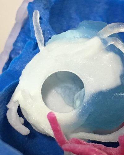
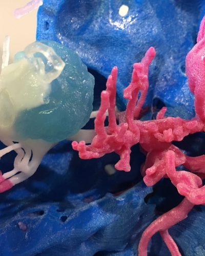
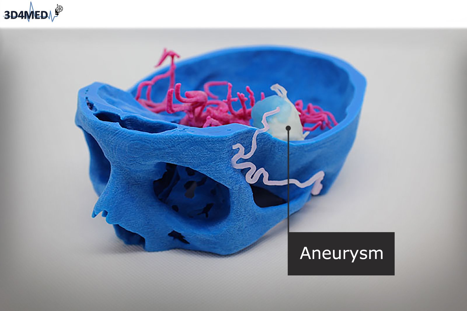
The 3D printed patient-specific model – including the artery aneurysm, its vascular proximal tract and the relative thrombus – was produced through Material Jetting Technology with different deformable photopolymer resins in order to assure the best mechanical adherence with the physiological reality; it was used to properly plan and simulate the surgical approach in an environment as much similar as possible to the operatory room’s one.


